
Breast cancer is one of the leading causes of death in Australian women, with one in seven women diagnosed with the disease in their lifetime. Stage 4 (metastatic) disease has a significantly lower survival rate than early-stage breast cancer.
Cancer cells spread from their initial site of growth at the primary tumour to secondary organs. It is vital to understand this aspect of disease progression as this will help in the development of treatments to reduce disease burden and mortality.
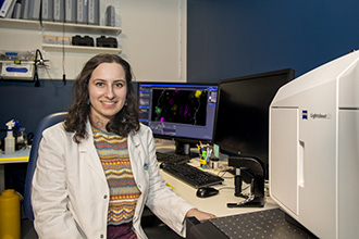
Sabrina Lewis, a PhD student supervised by Associate Professor Kelly Rogers and Dr Verena Wimmer, is using multiplexed, 3-dimensional imaging to combine information about cancer cell movement and blood vessel structure to better understand how tumour cells invade other tissues. She hopes this will lead to breakthroughs in novel cancer therapies.

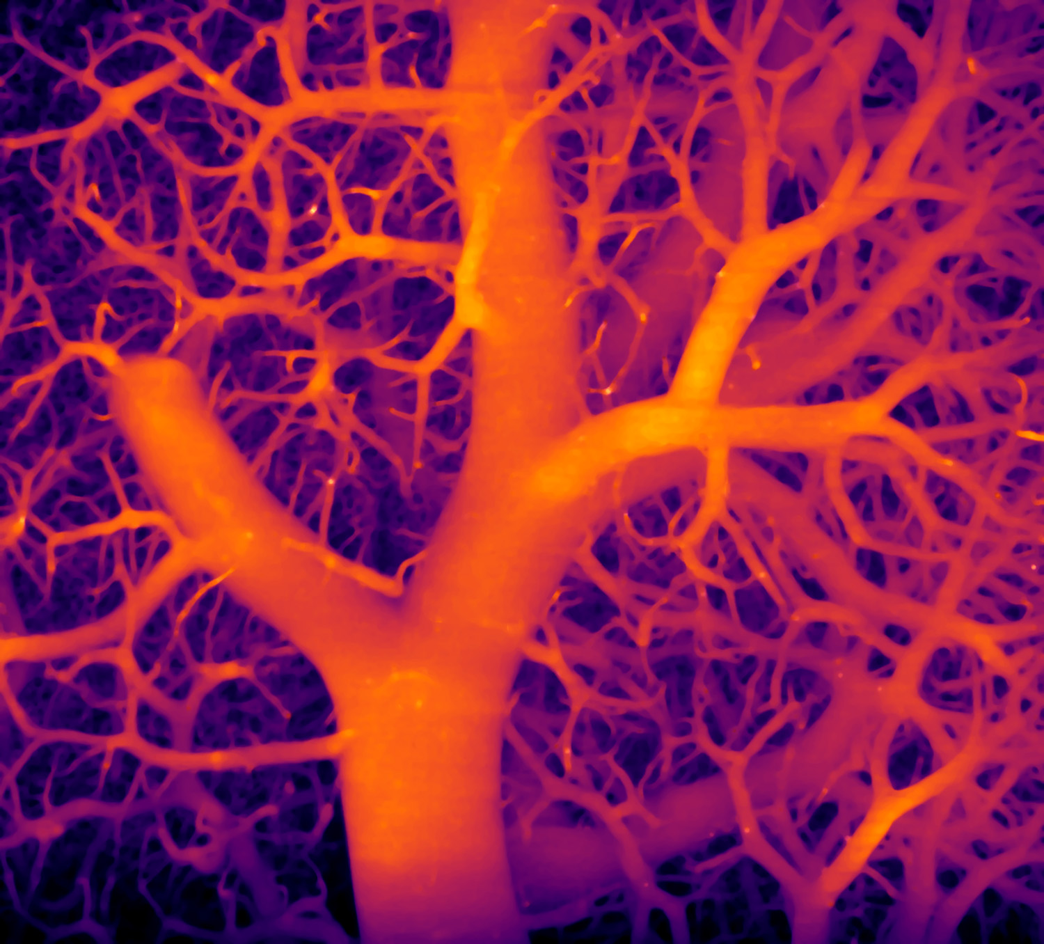
This tree-like structure is the complicated blood vessel network present in lung tissue. Ms Lewis is exploring new methods that enable cancer researchers to accurately pinpoint the location of cancer cells as they spread. Image: Sabrina Lewis
Cancer cell diversity affects efficacy of cancer therapy
Tumours are diverse tissue structures – this means they are composed of many different cell types (cancerous and non-cancerous cells). They can differ at the genetic, epigenetic, or phenotypic levels. High levels of cellular diversity within a tumour can influence the effectiveness of cancer therapeutics.
A 3-dimensional look at cancer
Limited research has been dedicated to understanding these challenging cancer cell dynamics in 3-dimensional tissue volumes. This has left a large gap in our knowledge of the variable interactions of cancer cells with other cell types as well as how cancerous cells spread through the body via the blood vessels.
Ms Lewis shines a light on these mysteries using lightsheet microscopy. This type of microscope specialises in imaging large-scale 3D tissue structures at high resolution. A semi-automated analysis pipeline is applied to the images that have been generated, which provides quantitative information about how these cancer cells interact within blood vessels.

