
Access to the Centre’s suite of imaging technologies has enabled WEHI researcher Dr Andre Samson to discover how the killer protein MLKL exerts its deadly job.

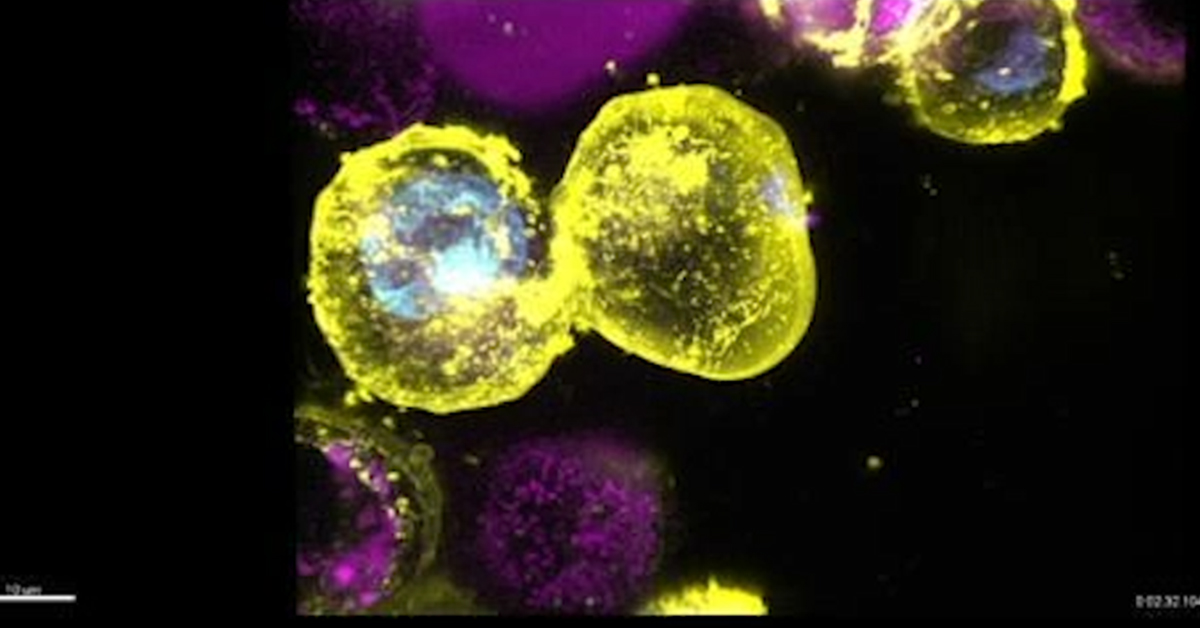
Cells dying by necroptosis, an inflammatory form of cell death which is driven by the protein MLKL
MLKL is a key player in the initiation of a specific type of cell death called necroptosis. This inflammatory cell death pathway is an important line of defence against infection but has also been linked to inflammatory diseases including dermatitis and inflammatory bowel diseases.

When Dr Samson started his study, it was known that MLKL travelled from cell’s cytosol the plasma membrane to execute necroptotic cell killing. However, the details how this happened remained elusive. Two years later, advanced imaging technologies have enabled Dr Samson and his colleagues to reveal important details about the mechanism of necroptotic cell death.
How does the killer travel to its execution site?
Dr Samson and the WEHI super-resolution specialist Dr Michael Mlodzianoski visualised MLKL moving into large cytosolic clusters to the plasma membrane where it then forms distinct hotspots in cells undergoing necroptosis.
This previously unrecognised process was monitored with the super-resolution imaging OMX/3D-SIM imaging technique, using staining of different cellular components in fixed cells to reveal MLKL’s exact cellular location. Visualisation of the plasma membrane by specific staining was game-changing in deciphering how MLKL killed cells, Dr Samson said.
How does MLKL kill cells?
The Centre for Dynamic Imaging’s lattice light sheet microscope enabled Dr Samson to watch in real time, as the plasma membrane began to bubble and blow up close to the MLKL ‘hotspots’. This ultimately led to the cell bursting – with the cellular contents spilling out and the cell deflating.
Cells falling like dominos
The team also revealed that necroptosis of one cell could trigger the domino-like death of neighbouring cells. This process was shown by time-lapse analysis using the Incucyte Live Cell Analysts System, an imaging technique that allows low-resolution tracking of large numbers of cells over time.
“I was fortunate to have access to a range of different imaging technologies, to allow me to visualise different aspects of MLKL’s role in necroptosis,” Dr Samson said. “These discoveries would not have been possible without the Centre for Dynamic Image’s suite of specialised technologies and the technical assistance provided by the centre’s imaging experts.”

