
Release of DNA from mitochondria has been implicated in a wide range of diseases, but precisely how the DNA escapes has never been explained.
Using a revolutionary new microscope, researchers have captured the moment when DNA is released from mitochondria during cell death.
The discovery has implications for understanding a wide range of autoimmune diseases.

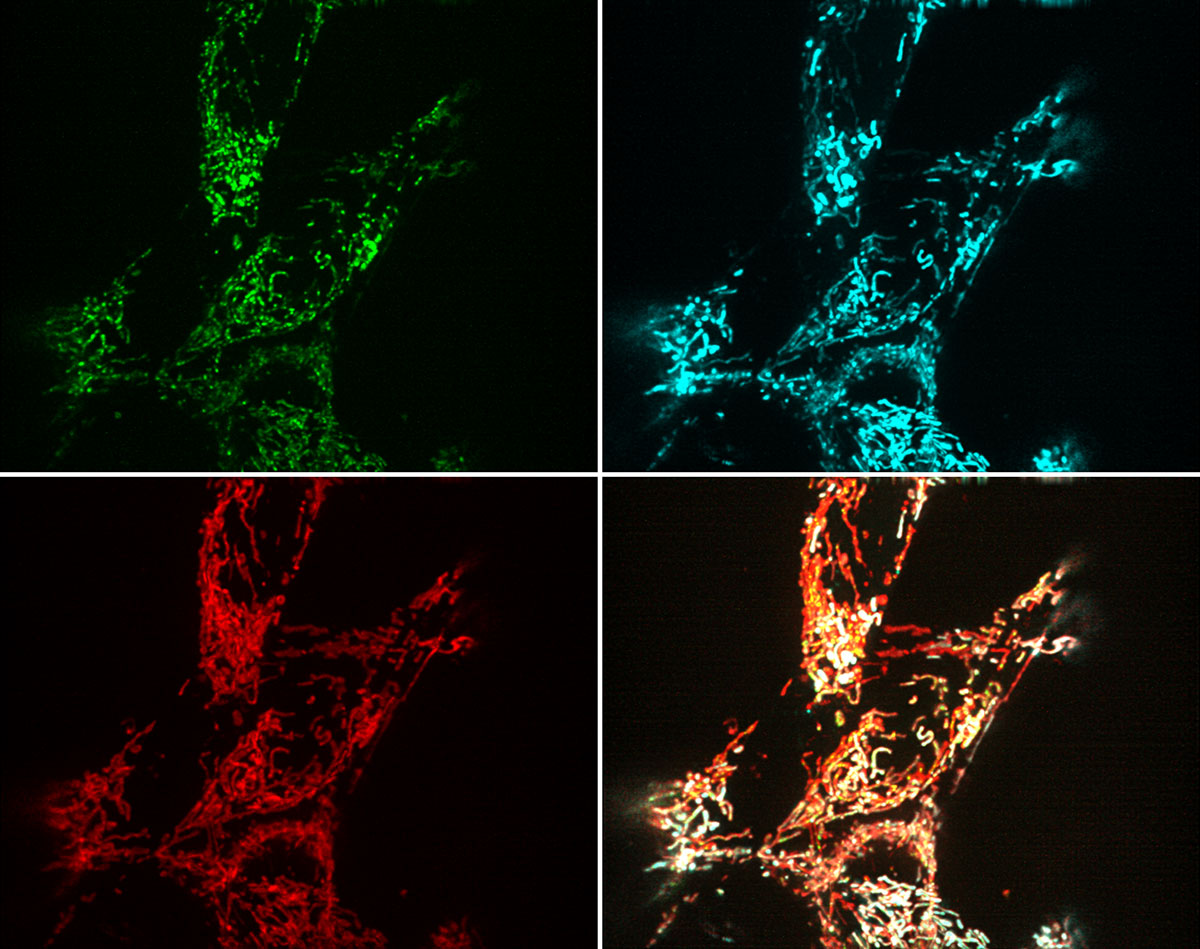
Mitochondrial DNA release during cell death, captured on the lattice light sheet. Credit: Kate McArthur, Niall Geoghegan

Mitochondrial DNA is linked to disease
Mitochondria are tiny structures within cells that generate energy to drive metabolic activities. The DNA inside mitochondria (mtDNA) has many similarities with bacterial DNA, and the body reacts to its presence outside the mitochondria as if under attack from invading pathogens. mtDNA release from the mitochondria is thought to contribute to autoimmune diseases such as arthritis and lupus.
Until recently, it was unclear how mtDNA escapes from mitochondria. Using conventional microscopes, scientists could not observe mtDNA release from mitochondria at sufficient resolution in real time to explain this mystery.
A revolution in microscopy

Dr Kate McArthur was studying mtDNA while working in the laboratories of Professor Guillaume Lessene and Professor Benjamin Kile.
Dr McArthur used the revolutionary new lattice light sheet microscope to examine mtDNA release during cell death. The lattice light sheet microscope is a new technique that allows scientists to watch the inner workings of living cells with unprecedented detail and in real time.
For her initial experiments, Dr McArthur travelled to the Howard Hughes Medical Institute Janelia Research Campus, US, where the lattice light sheet microscope was developed.
At the same time, scientists at the Centre for Dynamic Imaging were building the Institute’s own lattice light sheet microscope. It is the only custom-built microscope of its kind in Australia and was used in the final stages of Dr McArthur’s project.
mtDNA release from mitochondria captured for the first time
Using the lattice light sheet microscope, Dr McArthur was able to observe mtDNA release for the first time. She focused on cells undergoing programmed cell death, or apoptosis - a normal part of the body’s balancing act to control blood cell numbers.
Dr McArthur obtained spectacular images capturing the moment during apoptosis when mitochondria form a ‘hernia’ that balloons out of the mitochondria, expelling mtDNA into the rest of the cell.
Collateral damage leads to disease
Dr McArthur’s experiments show that mtDNA escape from the mitochondria is a form of collateral damage during apoptosis. There are normally mechanisms to rapidly clear apoptotic cells before the immune system is alerted. If this process isn’t properly controlled, however, the immune system can be triggered to drive pathological inflammation.
The finding will help researchers study the cause of autoimmune diseases, and ultimately find treatments for these conditions.