Lattice light sheet microscopy is the current state of the art for live cell imaging, enabling unprecedented 4D imaging capabilities.
Overview
Imaging biology in 4D has been a significant challenge in recent decades. The light used for imaging usually damages biological samples, making long term time-lapse microscopy extremely difficult.
Lattice light sheet microscopy is at the cutting edge of technology for live cell imaging because it is incredibly gentle on samples, allowing high resolution imaging over time.
This lattice light sheet was custom-built by staff at the Centre for Dynamic Imaging and was the first of its kind in Australia.
Examples of work
-
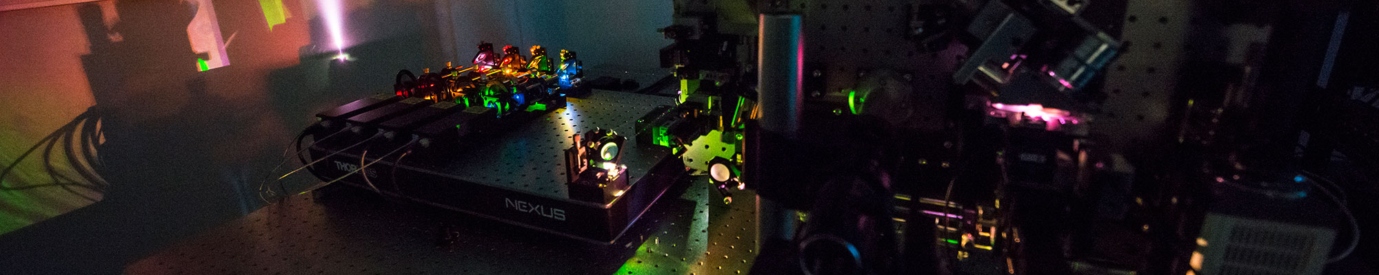
 The Institute's lattice light sheet microscope in the Centre for Dynamic Imaging
 -
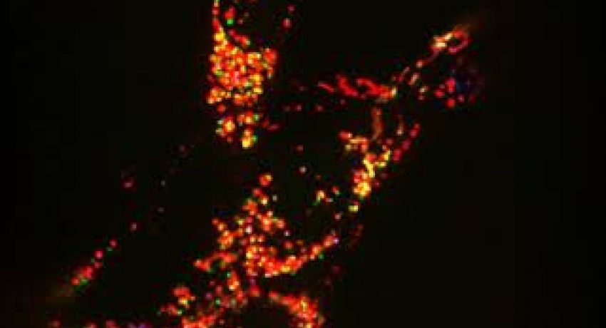
, mitochondria (green) and lysosomes (cyan). Credit: William Chang, Niall Geoghegan") Immune cell panel: nucleus (red), mitochondria (green) and lysosomes (cyan). Credit: William Chang, Niall Geoghegan
Immune cell panel: nucleus (red), mitochondria (green) and lysosomes (cyan). Credit: William Chang, Niall Geoghegan -
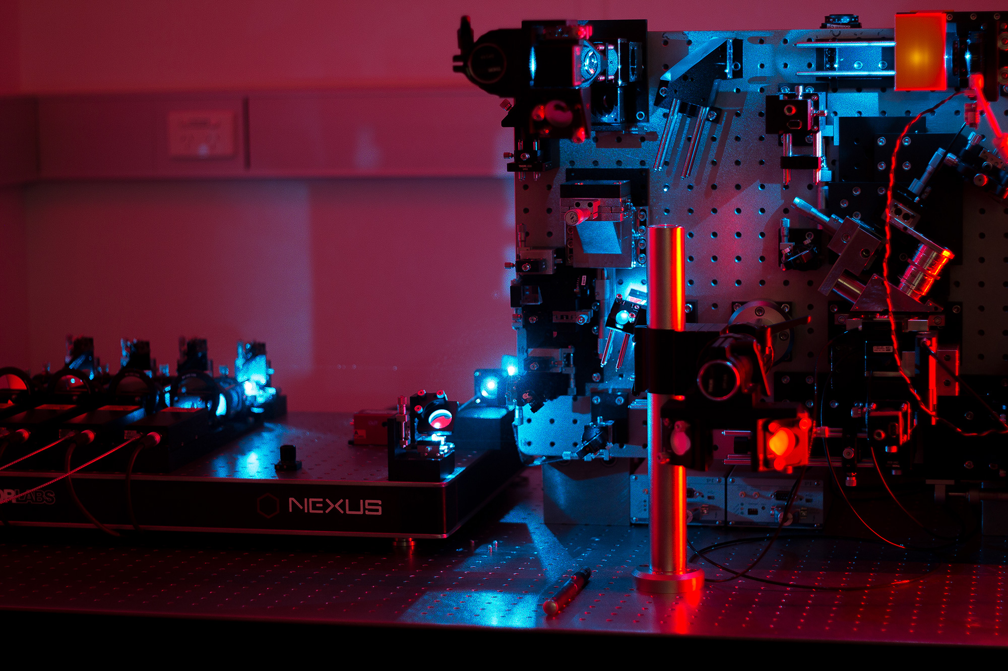
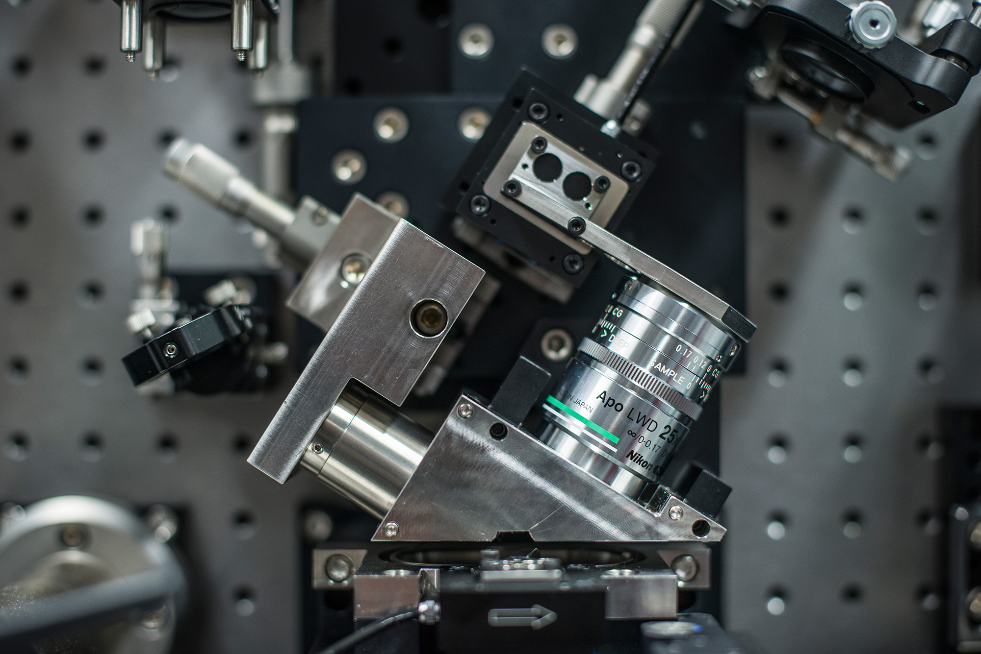
 Detailed view of the lattice light sheet microscope
 -
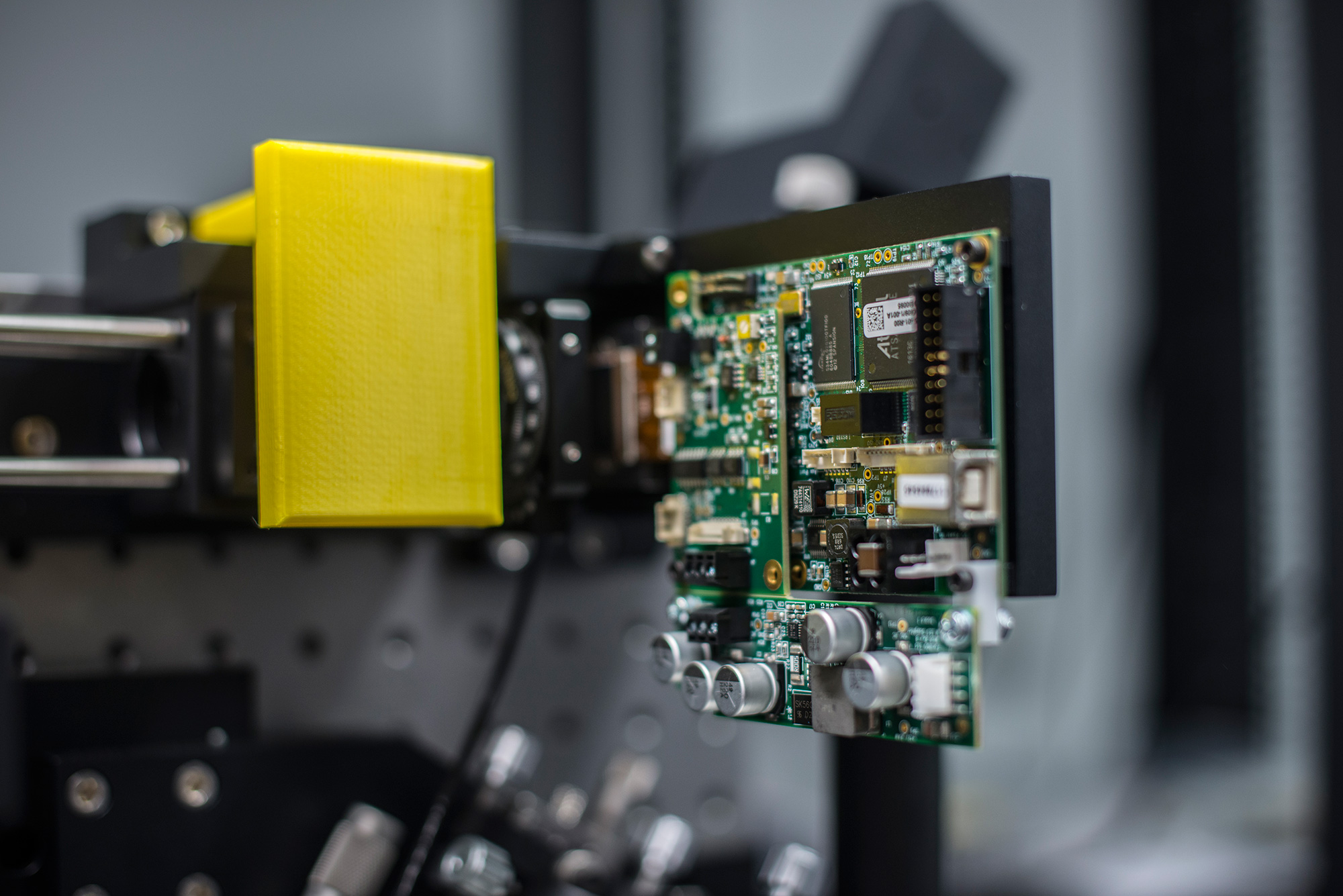
 Detail of the lattice light sheet microscope
 -
 Detail of the lattice light sheet microscope
-
The Institute's lattice light sheet microscope in the Centre for Dynamic Imaging
-
Immune cell panel: nucleus (red), mitochondria (green) and lysosomes (cyan). Credit: William Chang, Niall Geoghegan
-
Detailed view of the lattice light sheet microscope
-
Detail of the lattice light sheet microscope
-
Detail of the lattice light sheet microscope
Hardware
Objectives
Excitation objective
- Special optics 28.6x Magnification, 0.7 NA
Detection Objective
- Nikon Apo LWD Water dipping objective: 25x, 1.1 NA
Imaging characteristics
- 62x Magnification
- XY Resolution – 230 nm
- Z resolution – 370 nm
- Light sheet length – 10 µm – 100 µm
Light sources
- MPB fiber lasers – 488 nm, 561 nm, 589 nm and 642 nm
Detection source
- 2 x Hamamatsu Orca Flash 4 - V2
- 2048 x 2048 Pixels
- 6.5 µm x 6.5 µm pixel size
- 16 - bit
- 100 fps (At full frame)
- 82% QE (Peak at 560 nm)
Technological specifications (capabilities)
Available modalities
- Z-stack
- Time Series
- Multi-positions
Unique features
Lattice light sheet microscopy allows high resolution 4D imaging over many hours.
Limitations
- Complex sample geometry leads to unconventional sample mounting
- No transmitted light options
- No eye pieces
- Large quantities of data produced

